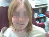
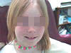
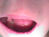
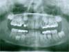
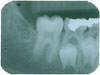
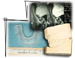
This ten-year-old child had a lesion on the right side of the mandible at the angle of the mandible.
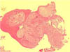
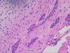
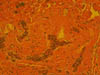
The lesion was identified as an ameloblastic fibro-odontoma, a recently defined entity, in which both ameloblastic fibroma and complex odontoma appear to be combined into one lesion. It has many clinical features in common with complex odontoma but differs significantly by having a greater potential for growth and local destruction.
Following biopsy it was removed completely.
|