The surgical pathology report described the specimen:
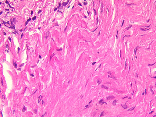
The tissue sections exhibit features of two distinctly different lesions. The larger portion of the specimen consists of moderately cellular spindle cell
proliferation amongst which there are numerous multinucleated giant cells. This portion of the specimen exhibits the characteristics of a giant cell granuloma. Other parts of the tissue consist of
highly vascular granulation tissue exhibiting an intense inflammatory cell infiltrate consisting largely of plasma cells, lymphocytes, and neutrophils and
exhibit the microscopic features of a periapical granuloma. The periphery of the specimen consists of relatively dense fibrous connective tissue and occasional small islands of vital bone. No evidence
of a cyst is seen in this material.