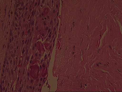
The surgical pathology report described the specimen:
The specimen consists of strips of dense fibrous connective tissue surfaced by a layer of
odontogenic epithelium containing occasional Rushton bodies. The cyst is non-keratinized but relatively uniform in thickness. The connective tissue wall contains
a patchy, mild, chronic inflammatory infiltrate and irregular fragments of proteinaceous material interpreted to
represent cyst contents are also present. No evidence of malignancy is present in these tissue sections.