
| The channel through which air enters the thorax (chest) is called the trachea, or windpipe. The trachea is a tube made of cartilage rings which keep the tube rigid and open for airflow. The rings are open at the back to acommodate muscles and membrane tissue which extend along the length of the windpipe. These muscles automatically tighten during exhalation and relax during inhalation, thus decreasing and increasing respectively the diameter of the trachea. |
| The trachea divides into two branches, called bronchi, which divide further into smaller and smaller vessels until there is a mass of spongy material (lungs), the walls of which are so thin that oxygen can pass through into the blood stream. |
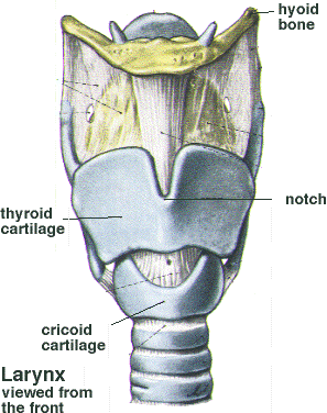
 At the top of the trachea is an organ called the larynx or voicebox, which plays a critical role in speech, singing, and breathing. The diagram on the left shows the larynx from the front. At the top of the trachea is an organ called the larynx or voicebox, which plays a critical role in speech, singing, and breathing. The diagram on the left shows the larynx from the front.
|
| The framework of the larynx contains two major cartilages: the cricoid cartilage, a ring-shaped cartilage on the top of the trachea; and the thyroid cartilage, which rests on the cricoid. You can feel the outline of the larynx by running your finger down the front of your neck. In fact, the protrusion in the thyroid cartilage at the notch shown in the diagram is commonly referred to as the "Adam's apple." |
| The larynx hangs from a U-shaped bone called the hyoid bone. The base of the tongue also attaches to the hyoid bone. The thyroid and cricoid cartilages are attached at the back in such a way that the thyroid is able to rock forward slightly on the cricoid. As explained below, this is an important feature used in manipulating the pitch of the voice. Above the vocal fold is a floppy cartilaginous tongue-shaped structure called the epiglottis. The epiglottis folds over the opening into the larynx when we swallow, which helps prevent material from getting into the lungs. |
 The second diagram shows a view of the larynx looking down from above. At the top edge of the back of the cricoid are two smaller cartilages called the arytenoids. As seen in the diagram, two vocal ligaments and two muscles are attached, one to each of the arytenoids and to the front of the thyroid cartilage. The ligaments and muscles stretch across the centre of the larynx forming a "V" when viewed from above. The muscles form the body of the vocal folds. The space between the vocal folds is called the glottis. There are many smaller muscles attached to the arytenoids which pull them apart during breathing (thus opening the airway more completely) and bring the arytenoids closer together during phonation in speech and singing.
|
| How does all of this work to produce a sound? It is important to realize that the vocal folds do NOT produce sound by vibrating like a string on a guitar or violin. During speech or singing, the vocal folds are brough closer together. As the air moves up from the lungs through the trachea and past the vocal folds, the folds open and close very quickly. This rapid pulsing of air passing through the vocal folds produces a sound which is then modified by the vocal tract (which extends from the folds to the lips of the mouth). The shape of the vocal tract can be changed by moving the tongue, jaw, palate, and lips. The sound is also amplified by the resonating nature of the voicebox itself. As well, the rocking action of the thyroid on the cricoid can cause the folds to stretch (producing higher pitches) or shorten and thicken (yielding lower pitches). This rocking is controlled by muscles attached to the thyroid and cricoid cartilages. |