Other parts of the brain involved in the control of movement.
The basal ganglia: anatomy, terminology and neuronal circuitry.
The term "basal ganglia," though anatomically unsatisfactory, is used extensively in neuroscience and clinical neurology.
BASAL
GANGLIA =
|
CORPUS STRIATUM
+ SUBSTANTIA NIGRA
+ SUBTHALAMIC NUCLEUS
|
The corpus striatum consists of the lentiform and caudate nuclei, which constitute the central grey matter of the telencephalon. The locations of these nuclei can be difficult to appreciate in 3 dimensions. The dissected specimens in the lab class are helpful, and it is essential also to understand what is seen in a
horizontal section
of the cerebral hemisphere at the level of the insula. The same preparations show the anatomy of the internal capsule.
The substantia nigra is easily visible in transverse sections through all levels of the midbrain. The subthalamic nucleus can be seen only in stained sections of the diencephalon.
Functionally, the corpus striatum has two parts:
|
STRIATUM =
|
CAUDATE NUCLEUS
+ PUTAMEN (This is the
lateral part of the
lentiform nucleus.)
|
|
PALLIDUM =
|
GLOBUS PALLIDUS (This is
the medial part of the
lentiform nucleus, and it is
subdivided into lateral [external]
and medial [internal] segments.)
|
Neostriatum is a synonym for striatum. Paleostriatum is a synonym for pallidum.
These terms are seldom used but you may encounter them in your reading.
The Crossman & Neary textbook includes the amygdala (= amygdaloid body) in the
basal ganglia. This is a traditonal anatomical assignation: the amygdala is a blob of grey matter,
comprising several nuclei, on the end of the tail of the caudate nucleus. Clinicians and basic scientists
who study the basal ganglia do not include the amygdala because its connections and functions are quite different.
Basal ganglia connections.
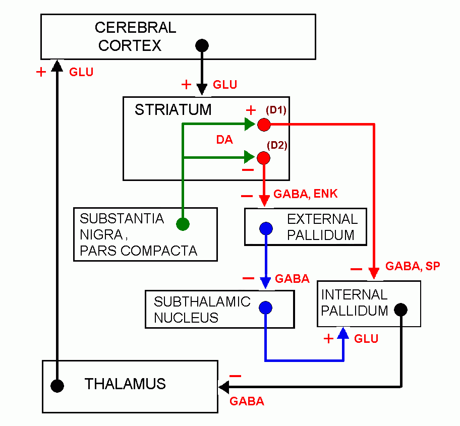
The connections of the basal ganglia and the associated neurotransmitters have been known for several years. Knowledge of this
circuitry can account for some of the clinical features of some of the diseases of the system. The information is simply presented
in this box-and-arrow diagram:

Follow the arrows to trace two pathways from the striatum to the motor cortex. The direct loop is the shorter pathway and has the net effect of stimulating the motor cortex.
The longer indirect loop, which goes through the external pallidum and subthalamic nucleus, inhibits the motor cortex.
The neurons of the striatum are continuously exposed to the action of dopamine delivered by the nigrostriatal tract.
This transmitter acts differently on the two types of striatal neurons. The cells that participate in the direct loop are
inhibited, whereas the striatal cells belonging to the indirect loop are stimulated. Both these actions of dopamine tend to
increase the excitation of the thalamus and motor cortex.
Basal ganglia diseases. These are called dyskinesias.
The commonest disease affecting the basal ganglia is Parkinson's disease, characterized by tremor at rest, rigidity
and bradykinesia ("poverty of movement"). The underlying cause is degeneration of the dopamine-producing neurons of the substantia nigra.
Reduced exposure of striatal neurons to dopamine may account for the bradykinesia of parkinsonism. Drugs used to treat schizophrenia block
dopamine receptors and can have parkinsonism-like side effects with conspicuous bradykinesia (tardive dyskinesia).
Other basal ganglia disorders include Huntington's disease and Wilson's disease, with degenerative changes in the corpus striatum
and unwanted movements of the limbs and face (chorea) that appear to be fragments of purposeful movements.
Athetoid movements are larger writhing movements of the limbs that complicate voluntary actions. Athetosis is seen typically in children;
it results from damage to the lentiform nucleus at the time of birth.
A vascular lesion in the subthalamic nucleus results in sudden onset of ceaseless large movements of the contralateral limbs, a syndrome known as
ballism or hemiballismus. It is attributable to loss of stimulation of the internal pallidum by the subthalamic nucleus, allowing
increased activity of the thalamocortical neurons.
_____________________________
Return to beginning of Anatomy 530a home page
Return to Anatomy 530a list of topics
