Return to beginning of Anatomy 530a home page
Return to Anatomy 530a list of topics
________________________________
Descending motor pathways and their disorders.
First, a bit about motor neurons and muscles. Some important new words and phrases are used in this summary. Look them up in a textbook, dictionary or other glossary if you don't already know them.
A motor neuron is a cell in the anterior horn (or in a motor nucleus of a cranial nerve) whose axon supplies the contractile fibres in skeletal muscles.
(There are 3 types of muscle tissue. Smooth muscle, composed of long cells that do not have cross-striations, occurs in blood vessels, internal organs and a few other places. It is supplied by neurons in autonomic ganglia. Striated muscle is composed of multinucleate fibres that have cross-striations because of the way their contractile proteins are aligned. There are two types of striated muscle. The heart is made of cardiac muscle, which has branched fibres with various other interesting structural features and is innervated by neurons in autonomic ganglia. All the ordinary muscles of the body, most of which pull on bones and move joints, have unbranched striated skeletal muscle fibres and are innervated by motor neurons.)
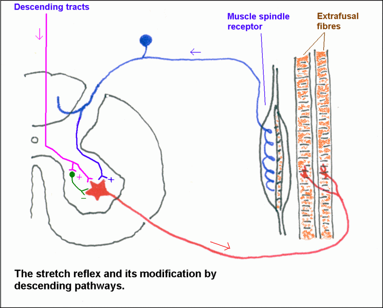
A motor unit comprises a group of extrafusal muscle fibers and the alpha motor neuron that innervates them. Gamma motor neurons supply the intrafusal fibers of muscle spindles. The term "lower motor neuron" is applied collectively to motor neurons.
| The stretch reflex requires sensory neurons that supply muscle spindles and motor neurons that supply the extrafusal fibres of the muscle. This reflex is ordinarily suppressed by the activity of descending pathways that end on motor neurons and (especially) on nearby interneurons. It can be elicited by suddenly lengthening a muscle, as when the tendon is tapped (jerk reflexes). |

|
The major descending pathways are the
vestibulospinal,
reticulospinal, and
corticospinal
(pyramidal) tracts.
The first of these is largely concerned with postural adjustments, and the last with voluntary movements. Most corticospinal fibers decussate at the caudal end of the medulla.
(Graduate students taking this course as Anatomy & Cell Biology 530a should read about the rubrospinal tract, which is part of a major motor pathway in mammals other than the great apes, man and woman.)
350a students: The textbook (Crossman & Neary) assumes there is a significant rubrospinal tract (eg diagram of spinocerebellar circuitry on p.122). This is wrong. Use the lecture and these web notes for guidance.
Diagram of the descending motor pathways
There is no such thing as an "upper motor neuron", but there is an entity called an upper motor neuron lesion. The latter phrase applies to clinical syndromes that result from destruction of the cell bodies or axons of the neurons in the major descending pathways.
An upper motor neuron lesion (such as transection of corticospinal and corticoreticular fibers in the internal capsule) causes spastic paralysis, with exaggerated stretch reflexes and the abnormal Babinski reflex (upgoing plantar reflex; down is normal. Atrophy is not a prominent feature, except when there is prolonged disuse.
Different causes of upper motor neuron paralysis (or paresis) cause different abnormalities.
Paresis = weakness, incomplete paralysis
For example:
LESION
TRACTS AFFECTED
FUNCTIONAL CONSEQUENCES
| Infarct involving either the motor areas of the cerebral cortex or the anterior limb of the internal capsule. |
Selective transection of corticospinal fibres.
Very rare; due to small lesion in either the medullary pyramid or the middle part of the cerebral peduncle. | Complete transection of the spinal cord. |
|
Corticospinal and corticoreticular projections are damaged.
The vestibular nuclei and vestibulospinal tract are intact. |
Corticospinal fibres are severed
Corticoreticular and reticulospinal projections are spared. | Corticospinal, reticulospinal and vestibulospinal fibres are severed. |
These positions are attributed to the action of the vestibulospinal tract. |
|
|
Corticobulbar fibers go to the motor nuclei of cranial nerves, in many cases bilaterally. It is assumed (but not known) that the parallel cortico-reticulo-bulbar projections are also bilateral.
Transection of these fibers in the internal capsule causes contralateral weakness of muscles of the lower half of the face and of the tongue, but not elsewhere in the head. This is because the lower motor neurons that supply the masticatory muscles, upper facial muscles, pharynx and larynx receive afferents from both cerebral hemispheres.
If a facial paralysis affects muscles acting on the eyebrow and forehead, the causative lesion involves the facial motor nucleus or fibres of the facial nerve. That is, it is a lower motor neuron lesion.
Click here for a diagram explaining why an upper motor neuron lesion affects only the lower face.
Motor areas of the cerebral cortex. Corticospinal, corticobulbar and corticoreticular fibres with motor functions are the axons of cells in four cortical areas.
You can look at the locations of these areas in a
picture showing functional cortical areas
These motor cortical areas project to one another and also to the spinal cord and those parts of the reticular formation that give rise to the reticulospinal tracts.
A chain of command (hierarchical projections) is recognized, beginning with the anterior frontal lobe cortex (thinking, predicting etc), passing through the cingulate, supplementary and premotor areas, then through the primary motor area.
In addition there are parallel projections of various cortical areas to the anterior frontal cortex and the various motor areas. These connections allow the motor areas to be influenced by sensory feedback.
The corticospinal and corticoreticular fibres originating in the cingulate, supplementary and premotor areas constitute descending projections parallel to that from the primary motor area.
The cortical areas are interconnected by association fibres: short ones that join adjacent areas, and long bundles that interconnect the different lobes.
Click here for a diagram of motor circuitry that emphasizes hierarchical and parallel cortical projections.
______________________________
________________________________
Return to beginning of Anatomy 530a home page
Return to Anatomy 530a list of topics