The spinal cord is shorter than the spinal canal in which it is suspended. Except in the neck, spinal cord segments are rostral to the corresponding vertebrae. The end of the adult spinal cord (the conus medullaris) is level with the 2nd lumbar vertebra.
Lumbar puncture: Cerebrospinal fluid can be sampled by a needle put into the subarachnoid space below the level of the conus medullaris.
The cross sectional area of the central gray matter indicates the number of neurons: largest for segments supplying limbs.
(Different levels)
The cross sectional area of the white matter decreases caudally because there are fewer descending and ascending fibers.
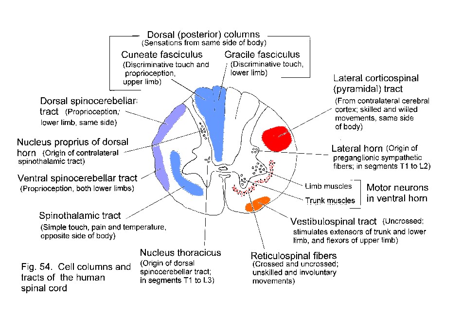
Motor neurons are in the ventral horn; sensory axons enter the dorsal horn and the dorsal funiculi. Preganglionic autonomic neurons are laterally placed, in segments T1-L2 and S2-S4.
Section of a thoracic segment of the spinal cord showing major groups of neurons in the grey matter, and positions of tracts in white matter.
Ascending tracts include the uncrossed gracile and cuneate fasciculi (from sensory ganglia) and the crossed spinothalamic tract (from the dorsal horn). These are concerned with different types of sensation.
Descending motor tracts include the uncrossed vestibulospinal and the crossed lateral corticospinal tract. Hypothalamospinal and some reticulospinal fibers influence autonomic functions.
Lesions in different parts of the spinal cord produce sensory and motor abnormalities appropriate to the functions of the tracts that have been transected. The segmental level of a lesion is indicated by the affected dermatomes and movements.
Lesion causing the Brown-Sequard syndrome
Can you determine what the motor and sensory deficits are, for parts of the body below the segmental level of the lesion?
_____________________________