History of Presenting Illness Physical Examination Basic Investigation Management Diagnosis Treatment Prognosis & Evidence-Based Info Summary Questions

In two weeks your patient returns feeling unwell, fatigued and experiencing shortness of breath and lightheadedness. Physical examination reveals patient is stable. No fever. BP: 100/70. HR: 70bpm. Prominent V wave in jugular pulsation and mild crackles in lung bases.

  • What do you see?
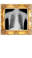
  • X-ray Results
  • What do YOU think is the problem?
  • Pacemaker Hemodynamics
    • The first 3 beats are ventricular paced with retrograde VA conduction resulting in a large “V” wave (may also be referred to as cannon A wave). This is likely atrial contraction against closed mitral - and tricuspid - valves
    • Another explanation could be Mitral Regurgitation from pacer-induced
      LV activation resulting in a V wave!
    • This is hemodynamic proof of pacemaker
      syndrome leading to elevated filling
      pressures and dyspnea!
  • Nuclear Scan
  • What would you do next?


The hemodynamic tracing shows LV and LA tracings.